Patients benefit most from time-saving Mohs surgery
USF dermatologist develops rapid immunostain technique
Word is spreading about a time-saving improvement to traditional Mohs surgery, a specialized outpatient method for treating certain skin cancers.
Since his break-through article (published last summer in the journal Dermatologic Surgery) revealed an improved technique that reduced the time for the immunostaining component of Mohs surgery from an hour to 19 minutes, USF’s Basil Cherpelis, MD, has seen a steady increase in the number of patients seeking Mohs surgery at the USF Health clinic. He has also seen an increase in the number of inquiries from other physicians around the country about the procedure.
“Physicians are calling from all over the U.S. to get more information about the protocol so they can offer this new technique in their own practices,” said Dr. Cherpelis, who is assistant professor of dermatology, chief of Dermatologic Surgery at USF, and associate director of the USF Dermatology Residency Program.

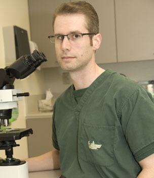
USF Mohs Surgeon Dr. Basil Cherpelis.
Mohs micrographic surgery was developed in the 1930s by Dr. Frederic Mohs as an innovative technique for completely removing certain skin cancers, such as basal and squamous cell carcinomas. Mohs surgery differs from other skin cancer treatments in that it allows for complete microscopic examination and mapping of the removed tissue to ensure removal of all cancerous cells. The result is a cure rate that is nearly 100 percent for skin cancers that have not been treated before.
Typically, patients needing Mohs surgery will spend several hours at the doctor’s office because of the time needed for staining each tissue sample to determine if cancer cells are present. The day is a series of repeated procedures, during which the surgeon removes a thin layer of skin at the site of the visible tumor along with a narrow rim of normal skin, stains and prepares the tissue sample, maps the locations of remaining cancer cells, and then returns to the patient to remove another section.
This continues until the tissue is cancer free. Sometimes special stains, called immunostains, are needed to visualize the cancer cells. Since the processing of immunostains takes an additional hour, in addition to the standard stains that are performed the entire procedure can easily take several hours, Dr. Cherpelis said. That is why patients and physicians alike are encouraged by Dr. Cherpelis’ rapid immunostain technique, which requires only 19 minutes for the tissue staining.
“I think the patients are the ones who are most excited about this reduced time, and that’s understandable,” Dr. Cherpelis said.
Currently, Mohs surgery is most beneficial to patients with basal and squamous cell carcinomas, but Dr. Cherpelis said that his practice is working to expand the procedure to include lentigo maligna melanoma.
“For lentigo maligna melanomas, it is difficult to identify the melanoma cells using standard stains and to determine distinguish between where the cancer starts and stops in the healthy tissue,” he said.
“It’s an area worthy of more research since melanomas are far more deadly.”
Dr. Cherpelis, who left private practice five years ago to join USF and open its first Mohs Surgery Unit, is a fellow of the American College of Mohs Surgery.
Titled “Innovative 19-Minute Rapid Cytokeratin Immunostaining of Nonmelanoma Skin Cancer in Mohs Micrographic Surgery,” the article was published in the July 2009 issue of Dermatologic Surgery, the journal of the American Society for Dermatologic Surgery. Co-authoring the article with Dr. Cherpelis is Logan Turner, MD, Sharron Ladd, BS, Frank Glass, MD, and Neil Fenske, MD.
For more information about USF’s Mohs Surgery program, visit USF Dermatology’s website or call 813-974-4744.
Story by Sarah A. Worth, USF Health Communications
Photos by Norman Weeks

