How are YOU put together?
USF Health anatomy exhibit featured at MOSI’s “The Amazing You”
Travel the journey from birth to end of life while exploring life stages and what it means to stay healthy. That’s just one lesson you walk away with after touring the interactive The Amazing You Exhibit at the Museum of Science and Industry (MOSI). This Spring, MOSI added a new phase to the ongoing exhibit that opened in 2008. The new wing features more fun educational displays on human life developmental stages and how wellness, stress, and disease management, among other conditions, can influence your physical health and well-being.

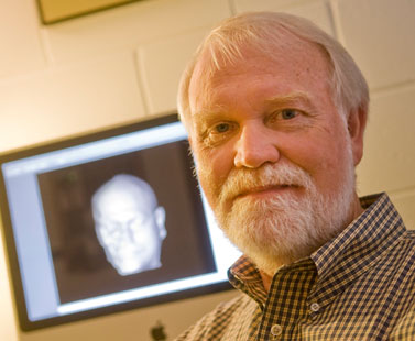
USF Health’s Dr. Don Hilbelink showcases a collection of human organs and a virtual human navigation system in the Museum of Science and Industry’s new wing.
In this new wing, Don Hilbelink, PhD, professor in the Department of Pathology & Cell Biology and Director of the USF Center for Human Morpho-Informatics Research (CHMR), showcases a collection of human organs and a virtual human navigation system allowing viewers to study human anatomy through 3-D medical imagery. The exhibit allows users to navigate through the body slice by slice to better understand details and structures of the human body and how we are built. Dr. Hilbelink’s lab also has provided examples of medical imaging scans like CT, MRI, and ultrasounds, which make it easy and fun for the public to understand the technology.

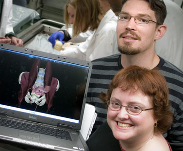
Summer Decker, MA, MS, graduate research associate in Pathology & Cell Biology, and Jonathan Ford, MS, graduate research assistant in Chemical and Biomedical Engineering, work with Dr. Hilbelink at the USF Center for Human Morpho-Informatics Research.
The CHMR has committed to keep the The Amazing You exhibit on the cutting edge by regularly updating the technology of the exhibits and the anatomical specimens that have been installed over the life of the exhibition (planned for 5 years). This ultimate 3-D experience offers a closer look at how our bodies work and how we are put together.
Watch the peel-away of a CT-scanned human head as USF Health’s Summer Decker describes the anatomy.
Story by Monica Matos, video by Klaus Herdocia, and photos by Eric Younghans, USF Health Communications

