USF Health isn’t usually called upon to scale the anatomy of a patient from human to animal, but the USF Radiology team has that expertise – and was ready when a Busch Gardens veterinarian reached out to schedule a CT scan for a young sloth with dental issues.
“When it comes to the specialty of radiology, Dr. Summer Decker and her team have been unbelievable,” said Dominique Keller, DVM, senior veterinarian at Busch Gardens Tampa Bay. In the past, USF Health helped the zoological park’s veterinary team with advanced diagnostic scans of penguins and wallabies, but this would be their first sloth.
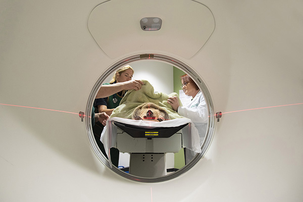
Kodiak, a 1-year-old, two-toed sloth, was seen by USF’s Radiology team for a CT scan to help determine treatment for an off-set jaw.
USF Health’s Summer Decker, PhD, assistant professor and director of imaging research in the Department of Radiology, holds several patented imaging techniques embracing 3D virtual and 3D printing technologies to visualize anatomy.
“We take pictures in slices and take all of those slices and push them back together to get the exact same patient in 3D,” Decker said. “You can essentially fly through the body.”
Kodiak, a 1-year-old, two-toed sloth, looked as though he was smiling when he arrived at USF in August 2017 for the CT scan. But, in fact, his “smile” was an off-set jaw, contributing to elongated teeth that prevented his mouth from closing properly and made eating difficult. The CT scan would help determine if Kodiak’s issue was a condition caused by his foot-sucking habit or a jaw deformity.

The CT scan and a 3D model of Kodiak’s head helped create prints that the team used to develop a dental treatment plan. Periodically filing down some areas of the sloth’s teeth allows his mouth to fully close.
First the radiation dose and scan protocol was modified to meet modified pediatric guidelines by USF CT technologist Joe Henry, and the images were reviewed by neuroradiologist Ryan Murtagh, MD, MBA, associate professor, Department of Radiology.
“Because of Kodiak’s size, he is basically a little kid, like a baby and has to be scanned differently than an adult,” Decker said.
The sloth’s unlikely interprofessional health care team included his veterinarian, a dentist, William Geyer DDS, two imaging research scientists and a radiologist. Before the craniofacial scan, they surrounded Kodiak and worked cautiously, avoiding his “razorblade sharp teeth.” While the images were generated, the team used normal sloth skulls from Busch Gardens’ skeletal reference collection to compare.
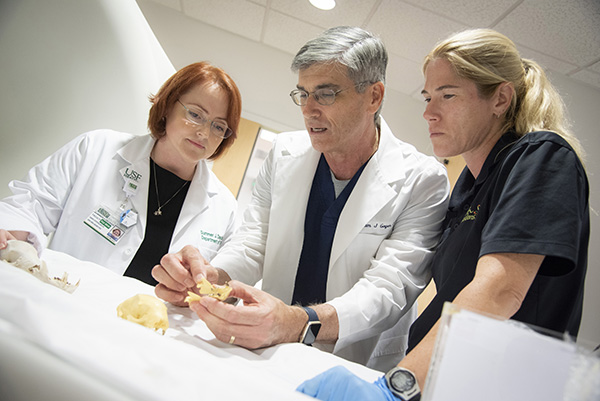
3D prints helped the interprofessional team visualize a treatment plan. From left, Summer Decker, PhD, assistant professor and director of imaging research in the USF Health Department of Radiology; Tampa dentist William Geyer, DDS; and Dominique Keller, DVM, senior veterinarian at Busch Gardens Tampa Bay.

Comparisons from normal sloth skulls shown with a 3D print from Kodiak’s scan.
“None of us had ever scanned a sloth before so we were unfamiliar with the anatomy. Using a reference like the normal skull allows us to determine where Kodiak’s differences are,” Decker said.
The 3D modelling analysis was operated by Jonathan Ford, PhD, biomedical engineer and assistant professor in the Division of Imaging Research at the Department of Radiology. No evidence of a jaw structural deformity was seen. But, Ford was able to virtually open and close Kodiak’s mouth to help identify the areas of jaw and teeth misalignment that led to Kodiak’s bite problem and show the changes that needed to be made to correct the issue.
“That’s why the CT is so nice” Dr. Keller said. “You can take an image, rotate it in three-dimensions and look exactly at what’s happening with him.”
In early October, the Busch Gardens team with Dr. Geyer returned to USF for a follow up. Decker’s team helped provide a treatment plan. A 3D print, made from the scan and 3D model, clearly showed where and how individual teeth would need shaving to correct the sloth’s bite.
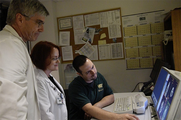
Dr. Geyer and USF Health’s Decker review digital models of Kodiak’s craniofacial scans with Joe Henry, USF CT technologist.
A couple weeks later, dental consultant Dr. Geyer shaved down Kodiak’s teeth at the Busch Gardens Animal Care Center with the whole team looking on. The procedure was simple and also a teaching moment for the small group of children who watched behind a glass wall.
“Will he be OK?” they asked.
Kodiak did well. However, his teeth will continue to grow and he’ll need similar dental treatments periodically for the rest of his life.
“This was very different from what we normally do,” said Decker, whose 8-year-old-self was fascinated to learn about sloths and jumped at the opportunity to “draw on all the different fields and combine that knowledge to create a solution.”
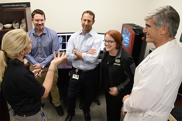
USF Health’s radiology team worked with a veterinarian and a dental consultant from Busch Gardens Tampa Bay to form the unlikely team. Members drew upon their different fields to create a solution.
The USF Radiology team thrives in USF Health’s academic clinical environment. Besides being just a call away from Dr. Keller, the clinical researchers have their hands in many other areas such as gynecology, neurosurgery, cardiothoracic surgery, plastic surgery, pathology and forensics, to name a few. Their expertise is routinely tapped by physicians and surgeons to help create “roadmaps” for treating patients, and they teach medical students and residents innovative, advanced imaging techniques.
“We need that collaboration. To me that’s the beauty of working near an academic medical institution, and USF is perfect for us,” Dr. Keller said.
A story about how the USF Health radiology doctors worked with the Busch Gardens Tampa Bay veterinary group to find a solution to Kodiak’s dental problem aired April 21 on the show Wildlife Docs.
-Video by Ryan Noone and Torie Doll, and photos by Ryan Noone, University Communications and Marketing
-Anne DeLotto Baier contributed to this story
